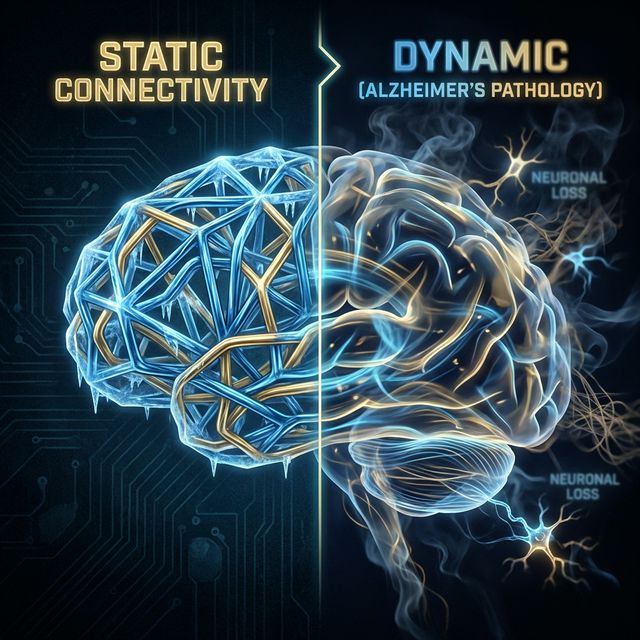
Functional connectivity (FC) has long been the gold standard for understanding how different brain regions communicate. In thousands of papers, researchers scan a brain for 10 minutes, calculate the average correlation between every pair of regions, and produce a “connectome.”
However, this approach suffers from a fundamental limitation: it assumes these connections are static over the course of scan. It collapses the rich, time-varying movie of brain activity into a single, frozen photograph.
My research in the MIND Lab challenges this assumption. By looking at Quasi-Periodic Patterns (QPPs)—recurring spatiotemporal events that drive much of the brain’s functional architecture—we are finding that the dynamics of the brain break down long before the static structure does.
The “Static” Limitation
Imagine trying to understand a symphony orchestra by looking at a long-exposure photograph taken over the entire concert.
- You would see that the violins sit near the cellos (topology).
- You would see that the percussion section is generally loud (amplitude).
- But you would miss the music itself. You wouldn’t know if they were playing Beethoven or Mozart. You wouldn’t hear the tempo changes or the call-and-response between sections.
Traditional static FC is that long-exposure photograph. In neurodegenerative diseases like Alzheimer’s, the “music”—the timing of neural communication—often falls out of sync long before the structural “wiring” physically disintegrates.
Study Design: TG2576 Mouse Model
In our recent work, we utilized resting-state fMRI (rsfMRI) to examine these dynamics in TG2576 mice.
- The Model: TG2576 is a well-established mouse model of amyloid pathology. These mice overexpress a mutant form of the human amyloid precursor protein (APP), leading to plaque deposition similar to human Alzheimer’s.
- The Method: Instead of just correlating regions, we used a pattern-finding algorithm to identify QPPs—repeating templates of brain-wide activity that last about 3 to 6 seconds in mice.
Key Findings: The “Blurring” of Networks
Our analysis revealed a striking dynamic failure in the Alzheimer’s mice that static analysis largely missed.
1. The Breakdown of Anti-Correlation
In a healthy brain (mouse or human), the Default Mode Network (DMN) and the Task-Positive Network (TPN) are natural antagonists. When you are focused on a task (TPN on), your internal monologue suppresses (DMN off). When you daydream (DMN on), your attention to the outside world fades (TPN off).
- Healthy Controls: We observed robust QPPs where the DMN and TPN were strongly anti-correlated. They fired in a precise, alternating rhythm.
- TG2576 Mice: This relationship collapsed. The DMN and TPN began to fire together, or their timing became “slushy.” The sharp dynamic boundary between “internal” and “external” attention was blurred.
2. The “Blurring” Effect
We visualized this as a “phase portrait” of brain activity. Healthy brains traverse a wide, structured state space—moving decisively between specific network configurations. The Alzheimer’s brains appeared “stuck” or “limited,” unable to fully switch states. The networks were dynamically blurring together.
3. Superior Classification
Crucially, when we trained a classifier to distinguish healthy mice from TG2576 mice:
- Static FC: Achieved moderate accuracy (~70%). The average connections were slightly weaker, but individual variability was high.
- Dynamic QPP Metrics: Achieved significantly higher accuracy (>85%). By measuring the strength and frequency of these QPPs, we could detect pathology much more reliably.
Why This Matters
This suggests that dynamic biomarkers could serve as reliable early warning signs.
- Early Detection: If dynamic coordination fails before plaques fully take over, we might be able to detect “at-risk” brains years earlier.
- Mechanism: It points to a failure of inhibitory control. The inability to shut down the DMN when the TPN comes online (and vice versa) suggests a breakdown in inter-neuron signaling that serves as the conductor of the orchestra.
By treating the brain as a dynamic system rather than a static graph, we can detect subtle network failures that traditional methods miss. We are moving from looking for “broken wires” to looking for “bad timing.”